Utilizing Light Touch to Assess Motor Cortex Inhibition in Muscle Weakness Associated with Pain
- muscleiq2
- May 17, 2025
- 8 min read

Introduction
Muscle weakness is a common clinical presentation in patients with musculoskeletal pain, often observed in conditions such as chronic low back pain (CLBP), rotator cuff tears (RCT), cervical radiculopathy, and medial collateral ligament tears (MCL). Emerging evidence suggests that this weakness is not solely due to peripheral muscle impairments but is significantly influenced by central nervous system mechanisms, particularly motor cortex inhibition. Pain-induced alterations in corticospinal excitability, characterized by increased short-interval intracortical inhibition (SICI) and reduced intracortical facilitation (ICF), can suppress motor output, leading to reduced muscle activation and force production. Recent studies highlight the potential of non-nociceptive somatosensory inputs, such as light touch, to modulate these inhibitory processes, offering a novel diagnostic tool for physical therapists to differentiate centrally mediated muscle weakness from peripheral causes.
This chapter explores the neurophysiological basis for using light touch over a painful body part to assess motor cortex inhibition as a contributor to muscle weakness. It provides a detailed, evidence-based protocol for physical therapists to implement this technique, supported by findings from 43 studies on pain, motor control, and somatosensory modulation. The chapter also discusses clinical implications, limitations, and future directions for integrating this approach into physical therapy practice.
Neurophysiological Basis for Light Touch Assessment
Pain and Motor Cortex Inhibition
Pain significantly alters motor cortex function, as demonstrated by transcranial magnetic stimulation (TMS) studies. In patients with chronic pain conditions, such as CLBP or RCT, motor-evoked potentials (MEPs) are reduced, indicating suppressed corticospinal excitability (Study 1, Study 9). This suppression is mediated by increased SICI, reflecting enhanced GABA_A receptor activity, and decreased ICF, linked to reduced glutamatergic facilitation (Study 4, Study 19). These changes are thought to be adaptive, restricting movement to protect the painful area from further injury (Study 19, Study 22). For example, a study on experimental muscle pain showed that pain increases SICI and reduces MEP amplitude, correlating with lower pain ratings when inhibition is stronger (Study 19). Similarly, in RCT patients, reduced MEP facilitation during voluntary deltoid activation suggests central reprogramming due to altered afferent input from the glenohumeral joint (Study 1).
This centrally mediated inhibition manifests clinically as muscle weakness, as seen in reduced peak torque in patients with musculoskeletal pain (Study 21). For instance, quadriceps strength in CLBP patients was significantly lower than in controls, with a lower central activation ratio indicating central, rather than peripheral, deficits (Study 22). These findings underscore that motor cortex inhibition is a key contributor to pain-related muscle weakness.
Role of Light Touch in Modulating Inhibition
Non-nociceptive somatosensory inputs, such as light touch, can modulate pain and motor cortex inhibition through sensory-motor integration. Tactile stimulation engages the primary somatosensory cortex (S1), which has strong monosynaptic connections to the primary motor cortex (M1) (Study 7). Studies demonstrate that homotopic tactile stimulation (e.g., to the painful area) triggers short-latency afferent inhibition (SAI), reducing corticospinal excitability, but also disinhibits local inhibitory circuits like SICI (Study 7, Study 8). This disinhibition can counteract pain-induced increases in SICI, potentially restoring motor output.
A key study on tactile-induced analgesia showed that light touch reduces pain perception at both spinal and cortical levels, with distinct spatiotemporal signatures in S1 (Study 23). Another study using a conditioning task found that tactile cues preceding painful stimuli diminish pain’s impact, suggesting competition between nociceptive and non-nociceptive inputs (Study 24). These findings indicate that light touch over a painful area may reduce motor cortex inhibition by modulating nociceptive drive and altering intracortical inhibitory networks, thereby facilitating muscle activation.
Clinical Rationale
The ability of light touch to modulate motor cortex inhibition provides a practical method for physical therapists to assess whether muscle weakness in a painful area is centrally mediated. By applying light touch and observing changes in muscle strength during the stimulation, therapists can infer the presence of motor cortex inhibition. An improvement in strength during tactile stimulation suggests that inhibition is reversible, pointing to a central mechanism, whereas no change may indicate peripheral or structural limitations. This approach leverages the neuroplasticity of sensory-motor pathways and aligns with evidence that somatosensory inputs can enhance corticomotor excitability (Study 5, Study 8).
Protocol for Assessing Motor Cortex Inhibition with Light Touch
Objective
To determine if muscle weakness in a painful body part is caused by motor cortex inhibition using light touch as a diagnostic tool.
Equipment
Dynamometer (handheld or isokinetic, e.g., Cybex) or manual muscle testing (MMT) equipment for strength assessment.
Patient Selection
Inclusion Criteria: Patients with musculoskeletal pain (e.g., CLBP, RCT, neck pain, knee pain) for >6 months, exhibiting muscle weakness in the painful area, and who have undergone conservative treatment (e.g., anti-inflammatory medication, physical therapy) (Study 1, Study 36).
Exclusion Criteria: Patients with neurological disorders (e.g., stroke, Parkinson’s), acute injuries (<6 months), or contraindications to strength testing (e.g., recent surgery, severe joint instability).
Procedure
Patient Preparation:
Explain the procedure to the patient, emphasizing that light touch is non-invasive and aims to assess the cause of muscle weakness.
Position the patient comfortably to access the painful body part (e.g., seated for shoulder, supine for low back).
Clean the skin over the painful area with an alcohol wipe to ensure consistent tactile stimulation.
Baseline Strength Assessment:
Select a target muscle associated with the painful area (e.g., deltoid for RCT, quadriceps for knee pain, lumbar multifidus for CLBP).
Measure baseline muscle strength using a dynamometer for objective torque output (e.g., peak torque in Nm/kg) or MMT (0–5 scale) for clinical settings (Study 21, Study 22).
Perform three trials, allowing 30 seconds rest between trials, and record the average strength value.
Note the patient’s pain level (0–10 numeric pain rating scale) during testing to account for pain interference.
Light Touch Application:
Identify the painful area (e.g., over the rotator cuff for RCT, L4–L5 region for CLBP).
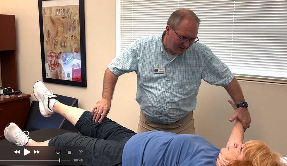
Using the middle three fingers (index, middle, ring) of the dominant hand, apply light, stationary touch to the painful area with gentle pressure (sufficient to indent the skin slightly but not cause discomfort). Maintain contact for 5 seconds. Ensure the touch is homotopic (directly over the painful site) to maximize S1-M1 interaction (Study 6, Study 7).
Monitor the patient to ensure the touch remains non-painful; adjust pressure if discomfort is reported.
Strength Assessment During Touch:
While maintaining the light touch (fingers stationary on the painful area), instruct the patient to perform the strength test for the target muscle (e.g., shoulder abduction for deltoid, knee extension for quadriceps).
Measure strength using the same method (dynamometer or MMT) as in the baseline, performing three trials during the 5-second touch period. If using a dynamometer, ensure rapid trials to fit within the timeframe; for MMT, assess resistance against manual pressure.
Record the average strength value and note any changes in pain level during testing.
Allow a 2-minute rest period, then repeat the light touch and strength assessment to confirm consistency of findings.
Control Condition (Optional):
To enhance diagnostic specificity, apply light touch with the middle three fingers to a heterotopic (non-painful) area (e.g., contralateral limb or adjacent non-painful region) for 5 seconds and repeat strength testing during the touch. Heterotopic stimulation is less likely to modulate pain-specific inhibition (Study 6).
Compare strength changes between homotopic and heterotopic conditions.
Interpretation of Results
• Positive Response (Central Inhibition Likely): An increase in muscle strength (e.g., higher torque or MMT score) during homotopic light touch suggests that motor cortex inhibition is contributing to weakness. This aligns with studies showing that tactile stimulation reduces SICI and enhances corticomotor excitability (Study 5, Study 8).
• No Change (Peripheral or Mixed Cause): If strength remains unchanged during light touch, weakness may be due to peripheral factors (e.g., muscle atrophy, structural damage) or irreversible central changes. Further diagnostic tests (e.g., EMG, imaging) are warranted.
• Pain Modulation: A reduction in pain rating during touch supports the involvement of tactile-induced analgesia, reinforcing the role of sensory-motor integration (Study 23).
Recording and Documentation
• Document baseline and during-touch strength values, pain ratings, and patient feedback.
• Note the specific muscle tested, touch application site, and any differences between homotopic and heterotopic conditions.
• Example table for documentation:
ConditionMuscleBaseline Strength (Nm or MMT)Strength During Touch (Nm or MMT)Pain Rating (0–10)HomotopicDeltoid2.0 Nm or 3/52.5 Nm or 4/55 → 3HeterotopicDeltoid2.0 Nm or 3/52.0 Nm or 3/55 → 5Clinical Implications
This light touch protocol offers a non-invasive, cost-effective method for physical therapists to assess the central contribution to muscle weakness in painful conditions. Identifying motor cortex inhibition as a cause can guide targeted interventions, such as sensory-motor training, aerobic exercise, or manual therapy, which have been shown to enhance corticomotor excitability and reduce inhibition (Study 5, Study 36, Study 39). For example, moderate-intensity aerobic exercise decreases SICI and improves motor learning, which could complement tactile interventions (Study 5). Additionally, this approach may inform rehabilitation by prioritizing strategies that address neuroplastic changes over purely strength-based exercises, which may be less effective in the presence of inhibition (Study 1).
Limitations and Considerations
• Variability in Response: Patient-specific factors, such as pain chronicity, psychological distress, or cortical reorganization, may influence the effectiveness of light touch (Study 1, Study 22). For instance, CLBP patients with high psychological distress showed lower central activation ratios (Study 22).
• Diagnostic Specificity: While light touch can indicate central inhibition, it does not rule out concurrent peripheral contributions. Complementary assessments (e.g., ultrasound, EMG) are recommended.
• Standardization: The pressure applied by the fingers must be consistent to ensure reliable results. Therapist training is essential to minimize variability.
• Contraindications: Avoid light touch in areas with hypersensitivity (e.g., allodynia) or acute inflammation, as it may exacerbate pain.
• Timing Constraints: Strength testing during the 5-second touch period requires rapid execution, which may be challenging for some dynamometer-based assessments.
Future Directions
Further research is needed to validate the stationary light touch protocol across diverse pain conditions and establish normative strength change thresholds for clinical significance. Integrating TMS or electroencephalography (EEG) with this protocol could provide direct measures of cortical excitability changes, enhancing diagnostic precision. Additionally, longitudinal studies should explore whether repeated light touch interventions can sustain reductions in motor cortex inhibition, potentially serving as a therapeutic tool. Investigating the role of subcortical regions in tactile modulation, as suggested by one study (Study 6), may also deepen our understanding of the underlying mechanisms.
Conclusion
Light touch over a painful body part, applied using the tester’s middle three fingers for 5 seconds, offers a promising diagnostic tool for physical therapists to assess motor cortex inhibition as a contributor to muscle weakness in musculoskeletal pain. By leveraging the modulatory effects of non-nociceptive somatosensory inputs on sensory-motor pathways, this simplified technique provides insights into central mechanisms of weakness, guiding personalized rehabilitation strategies. The protocol outlined in this chapter, grounded in robust neurophysiological evidence, is practical for clinical settings and aligns with the evolving understanding of pain’s impact on motor control. As research advances, this approach may become a cornerstone of physical therapy diagnostics, enhancing outcomes for patients with pain-related muscle dysfunction.
References
• Study 1: Chronic rotator cuff tear patients and motor cortex changes. Central neuromuscular dysfunction of the deltoid muscle in patients with chronic rotator cuff tears. https://jorthoptraumatol.springeropen.com/articles/10.1007/s10195-009-0061-7
• Study 4: Muscle pain and differential modulation of SICI and ICF. Muscle pain differentially modulates short interval intracortical inhibition and intracortical facilitation in primary motor cortex. https://www.jpain.org/article/S1526-5900(11)00870-4/fulltext
• Study 5: Aerobic exercise effects on motor learning and cortical excitability. Acute Aerobic Exercise at Different Intensities Modulates Motor Learning Performance and Cortical Excitability in Sedentary Individuals. https://www.eneuro.org/content/10/11/ENEURO.0182-23.2023
• Study 6: Tactile stimulation and afferent inhibition in M1. Modulation of Motor Cortical Inhibition and Facilitation by Touch Sensation from the Glabrous Skin of the Human Hand. https://www.eneuro.org/content/11/3/ENEURO.0410-23.2024
• Study 7: S1-M1 connections and motor control. The influence of sensory afferent input on local motor cortical excitatory circuitry in humans. https://www.ncbi.nlm.nih.gov/pmc/articles/PMC4386965/
• Study 8: Concussion, exercise, and cortical plasticity. Effects of Acute Aerobic Exercise on Motor Cortex Plasticity in Individuals With a Concussion History. https://uwspace.uwaterloo.ca/bitstream/handle/10012/18930/Khan_Madison.pdf?sequence=5&isAllowed=y
• Study 19: Pain-induced changes in MEP and TEP N45 peak. Alterations in cortical excitability during pain: A combined TMS-EEG Study. https://www.biorxiv.org/content/10.1101/2023.04.20.537735v2
• Study 21: Experimental muscle pain and central force inhibition. Inhibition of maximal voluntary contraction force by experimental muscle pain: A centrally mediated mechanism. https://onlinelibrary.wiley.com/doi/10.1002/mus.10225
• Study 22: Quadriceps strength and central activation in CLBP. Pain-Related Factors Contributing to Muscle Inhibition in Patients With Chronic Low Back Pain. https://journals.lww.com/clinicalpain/fulltext/2005/05000/pain_related_factors_contributing_to_muscle.6.aspx
• Study 23: Tactile-induced analgesia at spinal and cortical levels. A spatiotemporal signature of cortical pain relief by tactile stimulation: An MEG study. https://www.sciencedirect.com/science/article/abs/pii/S1053811916000951
• Study 24: Pain-tactile interaction in conditioning tasks. When touch predicts pain: predictive tactile cues modulate perceived intensity of painful stimulation independent of expectancy. https://www.degruyter.com/document/doi/10.1016/j.sjpain.2015.09.007/html
• Study 36: Strength training in CLBP. Effectiveness of a Group-Based Progressive Strength Training in Primary Care to Improve the Recurrence of Low Back Pain Exacerbations and Function: A Randomised Trial. https://www.mdpi.com/1660-4601/17/22/8326
• Study 39: Combined physiotherapy for shoulder pain. Which Multimodal Physiotherapy Treatment Is the Most Effective in People with Shoulder Pain? A Systematic Review and Meta-Analyses. https://mdpi-res.com/d_attachment/healthcare/healthcare-12-01234/article_deploy/healthcare-12-01234.pdf?version=1718886349


Comments