Fax #: 801-377-3697


Journal of Muscle IQ
Journal of Muscle IQ - Volume 10
August of 2026
Muscle Weakness Is Almost Always Inhibition
Find the nociception, restore the strength — the physiology behind it, the studies that confirm it, and seven demonstrations from the clinic.
by Chris Knudsen, DPT
Muscle IQ Physical Therapy
Orem, Utah · MuscleIQ.com
For practicing physical therapists.
The claim
When a muscle tests weak, the weakness is almost always muscle inhibition — the nervous system has turned the muscle's drive down — not the muscle mechanically failing. Something is sending nociceptive input, and that input inhibits the muscle. Find the nociception, reduce it, and the strength comes back. The MRI shows the surgeon where to put the scalpel; the weak muscle shows the physical therapist where to treat.
This is Inhibitory Driver Mapping: we are finding muscle inhibition, and then finding what is driving that muscle inhibition.
The established literature demonstrates that nociceptive input can inhibit motor output. Based on ten years of clinical observations, we also propose that this mechanism accounts for a much larger proportion of clinically observed weakness than is generally recognized.
This paper is about the weak muscle a physical therapist finds in an ordinary caseload; a musculoskeletal patient with an injured or irritable tissue, tested in the clinic. It is not a claim about every possible cause of weakness. A muscle can also be weak from causes this model does not cover: a myopathy, a motor neuron disease, a denervated muscle — where nothing turns back on because there is no drive left to restore. "Almost always" is meant within the population this paper is about, not as a statement about all weakness everywhere.
A second limit this paper has: Nothing here tells you which treatment to use. It is your call what treatment protocol to use; manual therapy, therapeutic exercise, modalities, etc. What this paper offers is the weak muscle test, and the three things it tells you: that the weakness is inhibition rather than a failed muscle; that inhibition you can reverse is inhibition you can rehabilitate; and where to aim the treatment.
This paper makes its case in this order:
-
How the nociceptive signal travels from tissue to muscle.
-
A weak muscle test is inhibition, not a failed muscle.
-
Where the weakness starts and how far it reaches.
-
Why damage and inhibition are not opposites, and why a tear or other structural finding does not equal the weakness — the imaging and strength research show both.
-
The physiology of how nociception weakens a muscle.
-
How a second nociceptive input restores strength to the first weakness.
-
The nociceptive input that can generate global weakening, and why it needs a condition to act on.
-
Seven demonstrations from the clinic.
-
What Inhibitory Driver Mapping lets you see.
-
What the research settles, what is our own clinical claim, and what future studies should try to resolve.
Figure 1
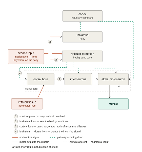
The machinery, from tissue to muscle
The path in summary. A nociceptor fires in irritated tissue — swollen, compressed, inflamed, or injured, whether or not the patient feels it as pain — and its signal enters the dorsal horn. From there it can reach the muscle by three routes of increasing length. Each route ultimately influences the alpha-motoneuron pool that drives the muscle.
-
The short loop. Straight onto interneurons in the cord, which act on the motoneuron directly. No brain involved.
-
The brainstem loop. Up to the reticular formation, which sends pathways back down that set the background tone of the muscles — working through those same spinal interneurons.
-
The cortical loop. Up to the cortex, then back down through the corticospinal tract, down to those same motoneurons. Nociceptive traffic arriving at the cortex can change the strength of the signal leaving the cortex to the motoneuron.
-
The fourth route acts on the signal rather than on the muscle: the brainstem sends fibers back down to the dorsal horn that dampen the incoming nociceptive traffic itself.
Three things arrive at the muscle’s motoneurons, from three different sources.
-
Voluntary command from the cortex, down the corticospinal tract. This is what creates voluntary movement.
-
Background drive from the reticulospinal system. It runs in the background whether or not the patient is doing anything, and is set by the state of the brainstem. This is the tone level.
-
Segmental input at the spinal cord — spindle afferents and the interneurons around them.
The alpha-motoneuron adds up whatever reaches it. How many of the muscle’s motoneurons fire, and how fast they fire, is what creates the muscle strength when tested.
A weak muscle test is inhibition, not a failed muscle
The simplest way to understand a weak muscle test is to ask a basic question:
Is the muscle actually incapable of producing force, or is the nervous system failing to fully activate it?
Experimental studies provide a remarkably direct answer.
When muscle pain was experimentally induced in healthy subjects, maximal voluntary knee-extension torque fell, but the muscle’s intrinsic contractile properties did not change when tested with twitch interpolation [2]. In other words, the muscle was still capable of producing force. What changed was the amount of force the nervous system allowed the subject to voluntarily produce.
That distinction is fundamental.
The muscle did not fail. The motor drive was reduced.
The study cannot determine whether that inhibition originated entirely in the spinal cord or at supraspinal levels. It does, however, establish the critical physiological point: nociceptive input can reduce voluntary muscle force without reducing the muscle’s ability to contract. This was an experimental pain study in healthy subjects rather than a clinical study of patients, but that is precisely what makes it useful here—it isolates the mechanism.
Two clinical studies demonstrate the same principle from the opposite direction: remove the nociceptive input, and strength can return almost immediately.
In fourteen patients with shoulder impingement—nine of whom had full-thickness rotator cuff tears—a subacromial local anesthetic injection was followed by repeat testing five minutes later. Thirteen of the fourteen patients became stronger on manual muscle testing, while isokinetic peak torque increased by an average of 48% [30]. Importantly, the strength increase did not differ according to whether a rotator cuff tear was present.
The muscle did not grow in five minutes.
The tendon did not heal in five minutes.
The input changed, and the output changed.
That is what inhibition looks like.
A second clinical study found the same phenomenon in the knee. In sixteen patients with chronic arthritis and clinically active synovitis, draining the knee joint produced an immediate increase in quadriceps peak torque (p = 0.004) [31]. After corticosteroid treatment, torque increased by an average of 25% over fifteen days. The immediate improvement cannot reasonably be attributed to muscle hypertrophy. Again, changing the nociceptive environment produced a rapid change in motor output.
These studies are small and have methodological limitations. They do not prove that every clinically observed case of weakness is caused by nociception. They demonstrate something more specific and important: a painful or irritated musculoskeletal structure can suppress voluntary motor output, and reducing that input can restore force without changing the muscle itself.
This distinction matters clinically because a manual muscle test measures output, not muscle integrity.
When a patient cannot generate normal force, the test does not tell us by itself that the muscle is damaged, torn, atrophied, or incapable of contracting. It tells us that the nervous system is not producing normal motor output through that muscle at that moment.
In the ordinary musculoskeletal patient seen in outpatient physical therapy, that difference changes how the finding should be interpreted.
A weak test should first make us ask, “What is inhibiting this muscle?” rather than assuming, “What is wrong with this muscle?”
Our clinical experience is consistent with this model. In the large majority of our patients, a weak muscle can show increased strength within minutes when the inhibitory driver is reduced or instantly (temporarily) when a competing nociceptive input is introduced. A muscle that can return to normal force in minutes or seconds did not lose its contractile capacity in the first place. Its output was being inhibited.
This is our clinical observation, not a controlled clinical trial. The research establishes that nociceptive input can inhibit motor output and that reducing nociceptive input can rapidly restore strength. Our clinical claim is that this mechanism accounts for a much larger proportion of the weakness encountered in ordinary outpatient musculoskeletal practice than is generally recognized.
That is the distinction this paper is proposing:
The weak muscle test is not necessarily evidence of a failed muscle. In the typical musculoskeletal patient, it is often evidence of an inhibited muscle.
Global Inhibition: Where weakness starts, and how far it reaches
Arthrogenic muscle inhibition is well established: injury, swelling, or irritation of a joint can reduce voluntary activation of the muscles surrounding that joint. A recent review notes that this inhibition can persist even after technically successful joint replacement, limiting recovery despite the structural problem having been corrected [29]. The joint can be repaired, yet the muscle still does not fully turn on.
The phenomenon is common. Among 300 consecutive patients with acute anterior cruciate ligament injuries, 56.7% demonstrated clinically detectable quadriceps inhibition before surgery [32]. A review of three common knee conditions—ACL injury, anterior knee pain, and osteoarthritis—found changes at multiple levels of the motor system, including the motor cortex, corticospinal tract, and spinal motoneuron pool. Across these conditions, altered excitability, reduced voluntary activation, and reduced force were consistently reported [33].
So there is little controversy about the basic phenomenon:
A musculoskeletal injury can inhibit motor output.
The more interesting question is how far that inhibition can reach.
The traditional concept of arthrogenic muscle inhibition focuses on the muscles surrounding the injured joint. That makes intuitive sense: the knee is irritated, so the quadriceps is inhibited. But that definition describes where the phenomenon was first observed; it does not establish that the inhibition must remain local.
Some evidence suggests otherwise.
In an experimental knee-pain study, tibialis anterior activity was also measured as a muscle outside the primary muscle group being tested. Tibialis anterior did not show the same change as the quadriceps [40]. That finding is important because it shows that inhibition is not automatically global. A nociceptive input does not simply turn down every muscle in the body.
But other evidence points toward a much wider reach.
In 107 patients with knee osteoarthritis, Simis and colleagues found that greater cartilage damage was associated with greater motor inhibition measured in a hand muscle, far from the affected knee [28]. The relationship was seen across two measures of cortical inhibition. Interestingly, reported pain did not follow the same relationship; the remote inhibition tracked the condition of the knee rather than simply the patient’s reported pain. The study was cross-sectional, and the authors interpreted the increased inhibition as a possible compensatory response. It therefore does not establish the mechanism I am proposing. But it does establish an important observation:
A pathological condition in one joint can be associated with altered motor inhibition in a muscle that is nowhere near that joint.
That observation raises a physiological question. How could a nociceptive input from one part of the body influence motor output somewhere else?
The brainstem provides a plausible answer.
The reticular formation contains descending systems that influence the excitability of spinal motor circuits throughout the body. Experimental stimulation studies in decerebrate cats have demonstrated opposing regions within the reticular formation: stimulation of some regions suppressed muscle tone bilaterally, while stimulation of neighboring regions increased it [45]. The effect could persist beyond the period of stimulation. The precise location of these inhibitory and facilitatory regions has differed between laboratories, so their exact anatomical boundaries remain unsettled. More importantly for this paper, those experiments did not apply nociceptive input. They demonstrate the existence of a system capable of changing muscle tone broadly; they do not by themselves prove that nociception recruits that system [45].
Our clinical observations suggest a sequence that may connect these pieces.
At rest, a persistent nociceptive source can produce local inhibition. The muscle closest to the irritated structure is weak.
Then the patient uses that inhibited region. A straight-leg raise, a lunge, or movement into a painful direction can increase nociceptive input. In our clinical testing, that increase is sometimes accompanied by weakness appearing in muscles that are not themselves injured.
This is the phenomenon we call global inhibition.
The proposed mechanism is that additional nociceptive input reaches brainstem circuits capable of altering descending motor drive. Rather than inhibiting only the muscles surrounding the original injury, the system can reduce the background excitability of motor pools elsewhere in the body. A muscle with no local pathology can therefore test weak because the descending drive to that muscle has been reduced.
The distinction is important:
Local inhibition asks, “Why is the muscle next to the injured tissue weak?”
Global inhibition asks, “Why did a muscle with nothing obviously wrong with it just become weak?”
Our two-stage clinical sequence is a clinical observation, not an established finding in the literature. We know of no study that directly compares the same nociceptive source at rest versus during active loading while simultaneously measuring strength in a muscle remote from the source.
That is precisely the gap our clinical model addresses.
The existing research establishes that musculoskeletal pathology can inhibit voluntary motor output, that inhibition can occur at multiple levels of the motor system, and that motor inhibition can sometimes be detected in muscles remote from the pathological joint [28,29,32,33]. The proposed extension is that a sufficiently strong or additional nociceptive input may recruit descending brainstem mechanisms that temporarily reduce motor output beyond the original region.
If that is correct, a weak muscle test does not necessarily tell us where the problem is.
It tells us where the inhibition is being expressed.
That is why the next question should not simply be, “What is wrong with this muscle?”
It should be:
“Where is the inhibitory signal coming from?”
What Can Generate the Global Inhibition — and Why It Needs a Condition to Act On
The first input has to be genuinely nociceptive. But the source of that input can be much broader than a palpable, tender lesion. Many ordinary stimuli can generate nociceptive input when they act on an underlying condition: pressure on a tender point, loading or using an injured joint, holding the spine in flexion, turning a painful neck, sustaining a gaze in one direction, or even exposure to bright light.
The important point is that none of these stimuli is inherently nociceptive. They become nociceptive because of the condition on which they act. A lunge is not inherently nociceptive; it can become one when performed on an injured or irritable knee. Spinal flexion is not inherently nociceptive; it can become one when performed in the presence of an underlying low-back disorder. Bright light is not inherently nociceptive; it can become one in a person with photophobia. Sustained gaze to one side is not inherently nociceptive; it can become one when an underlying cervical injury makes that input provocative.
This distinction is important because it provides a testable prediction: the same stimulus should produce very different effects in people with and without the underlying condition. The published eye-movement data illustrate this pattern. Eye-movement testing provoked symptoms in roughly one-third to two-fifths of concussed adolescent athletes, but in no more than 9% of healthy controls [49]. The eye movement itself is not the pathology. The difference is the condition on which the stimulus acts.
The condition also does not necessarily have to be consciously experienced as pain. Nociceptive input can exist below the threshold of conscious pain perception and still influence motor output, as demonstrated by the spinal finding discussed earlier [11]. Pain is therefore not required for a tissue condition to provide the input that drives inhibition.
Named diagnoses belong on the same list. A meniscus tear, a surgical incision with hardware in place, an L4–L5 disc herniation, cervical stenosis, or lateral epicondylitis can each provide a source of abnormal sensory input capable of influencing the nervous system. The relevant feature is not whether the patient describes the structure as painful at that moment. The relevant feature is that a physical condition exists that can alter the sensory input reaching the nervous system.
The healthy-subject evidence provides the other half of the argument. When the visual system was activated in healthy people who did not have photophobia, light exposure did not produce a nociceptive response [19]. That is exactly what the model predicts: without an underlying condition capable of converting the stimulus into nociceptive input, there is no nociceptive signal—and therefore no reason for the global inhibitory response to occur.
The stimulus is not the inhibitor. The condition makes the stimulus nociceptive; the nociceptive input then drives the inhibition.
Damage and inhibition are not opposites — and why the tear does not equal the weakness
At the beginning of the paper we laid out the main point of this paper: weakness is almost always muscle inhibition — the nervous system has turned the muscle’s drive down — not the muscle mechanically failing.
None of that means damaged tissue has little to do with weakness, as if the inhibition arrived out of nowhere. Often the inhibition is triggered by damaged tissue. The tissue generates the nociceptive signal, and that signal is what turns the muscle down. A rotator cuff tear, a torn meniscus, a herniated disc — any of these can be sitting right there on the image and be the driver of the weakness, not by subtracting contractile tissue but by what message it is sending. The knee osteoarthritis patients above make the point: worse cartilage damage went with more motor inhibition, on two separate measures [28] — an association across patients, not a change measured within one. Damage and inhibition rose together rather than standing in for one another.
The important question is not whether the weakness belongs to the tear or to the inhibition; it is whether the muscle can be turned back on. If it can, the weakness was inhibition, whatever the tissue looks like.
The mechanics. A tear removes some contractile tissue, but as long as there is enough intact muscle and enough surrounding synergists to compensate, that loss is often not enough to move the strength test on its own — once the irritation is reduced. Much of the weakness you measure can be inhibition rather than missing tissue — which is what the anesthetic and drainage studies above put numbers on [30, 31].
The scan does not match the symptom. Structural findings are common in people with no pain at all. In 230 knees of pain-free adults, 97% had at least one structural abnormality on MRI, including meniscal tears in 30% and cartilage abnormalities in 62% — with 41% showing moderate-to-severe lesions and 31% severe ones [3]. Elite volleyball players with no shoulder pain showed abnormal shoulder MRIs across the board, including partial rotator cuff tears [4] (a small study). And baseline lumbar MRI did not predict who would develop low-back pain over the following seven years [5]. A finding on a scan is common, and it is not the same thing as a symptom.
Where strength itself was measured, the tear does not track it as cleanly as you would expect — pain does. In 891 shoulders of women aged 64–87, rotator cuff tears of all sizes had no isolated effect on strength in those under 70, while age, pain and testing the non-dominant arm each reduced it. In the over-70s, full-thickness tears did reduce strength — by roughly 30% for small and 40% for large tears [6]. Age matters, and we are not saying a tear never costs strength. However, patients with tears severe enough to require surgery tested near-normal on manual muscle testing before repair — external rotation graded 4.4 out of 5 in traumatic tears and 4.5 in atraumatic ones [7]. Over 15 years, unrepaired tears grew from a mean of 16.2 mm to 31.6 mm in the anterior-posterior direction — close to double — while the strength difference in favor of repair was 1.8 kg and did not reach statistical significance [8]. And in isokinetic testing before and after repair, pain correlated with the supraspinatus’s initial peak torque while fatty infiltration grade correlated with nothing — though tear size did correlate with preoperative initial peak torque, so size is not irrelevant [9].
The same split shows up in surgical populations. In 61 patients scheduled for shoulder surgery, marked weakness in abduction and external rotation was recorded across every tear size, including in the eleven patients found at surgery to have no tear at all — though in that same study one deficit did track tear size: abduction strength at 10° of abduction, where 20 of 25 patients with large or massive tears lost more than half their strength against the other side, compared with 1 of 11 with no tear [34]. Every other strength and range-of-motion measure in it was unaffected by tear size. And a Cochrane review of physical examination for lumbar disc herniation concluded that most individual tests, muscle weakness among them, perform poorly when used on their own; note that nearly all of its studies came from surgical populations, where herniation is far more common than in an outpatient caseload [35].
Injured tissue does not equal muscle weakness. The scan is a data point, not a verdict.
Why nociception turns a muscle down
This is the physiology under the whole model. The key distinction is simple: nociception is not the same thing as pain. A nociceptor can fire when tissue is irritated, swollen, compressed, inflamed, or injured without the person consciously experiencing pain. The nervous system does not have to wait for pain to be felt before it responds to the incoming signal.
That distinction matters because the motor effect of nociception can be demonstrated without pain and without the brain being involved at all.
In the classic cat experiments cited here, the preparations were decapitated and high-spinal — there was no brain in the circuit and therefore no possibility of conscious pain. Nociceptive afferents were activated while researchers recorded the response of alpha-motoneurons. Rather than facilitating the muscle’s normal stretch-reflex input, nociceptive conditioning inhibited the monosynaptic input from group Ia muscle-spindle afferents to the alpha-motoneurons [11]. In other words, nociceptive input can directly reduce the neural signal reaching the motor neuron at the spinal level. The motor consequence of nociception does not require the signal to be felt.
Human evidence shows the same separation between pain and inhibition. After medial meniscectomy, quadriceps inhibition was measured following different amounts of local anesthetic injected into the knee. The lower anesthetic dose reduced pain without reducing the measured inhibition, while the higher dose reduced both. More importantly, three to four days after surgery, the electrical activity associated with maximal quadriceps effort remained markedly depressed — a median 75% below each person’s preoperative value — even though pain was mild or absent; at ten to fifteen days, inhibition was still about 35% with little or no pain. The authors concluded that the inhibition was not simply a consequence of perceived pain, but was due, at least in part, to stimuli arising from the knee [36]. The joint can therefore continue to inhibit motor output even when the patient is no longer experiencing significant pain.
There is also evidence that sensory input can inhibit motor output without pain being the relevant signal at all. During fatiguing exercise, group III/IV muscle afferents — sensory fibers that report the mechanical and metabolic state of active muscle — contribute to the fall in motor-cortex excitability. When those afferents were blocked with intrathecal fentanyl, the normal fall in motor-cortex excitability did not occur [39]. The authors attributed the effect to these sensory afferents facilitating inhibitory cortical interneurons using GABA. The nervous system can therefore reduce motor drive in response to incoming sensory information even when pain is not the conscious experience.
The same principle can be seen higher in the nervous system. Experimental pain changes the excitability of the motor system, including the corticospinal pathway. A systematic review and meta-analysis of 49 studies found that experimentally induced pain reduced corticomotor excitability when the pain lasted from milliseconds to hours, with large effects for the shortest-duration pain and moderate effects for pain lasting minutes to hours [10]. The literature is not uniform across every muscle, pain duration, or experimental condition, but the overall finding is clear: nociceptive pain can reduce the nervous system’s capacity to drive muscle.
The important point is that this inhibition is not necessarily confined to the muscle or body part receiving the nociceptive input.
A painful stimulus applied to one hand reduced motor-cortex output bilaterally [12]. Experimental pain applied to the knee interfered with motor-cortex responses during a hand motor task just as pain applied to the finger did [13]. Patients with chronic low-back pain also showed altered corticomotor excitability measured at a hand muscle, even though the nociceptive source was in the back [14]. These findings demonstrate that the motor consequences of nociception can extend beyond the tissues generating the signal.
This provides an important physiological explanation for a clinical observation that otherwise seems strange: a nociceptive source in one part of the body can be associated with reduced motor output in a muscle somewhere else.
There are several possible levels at which this reduction in motor output can occur. Nociceptive afferents can interact with motor circuits in the spinal cord [11]. Nociceptive input can alter brainstem and descending motor systems. And experimental pain can alter cortical excitability and corticospinal output [10, 12–14]. These are not competing explanations. They are different points along the same sensory-to-motor system.
The clinical model proposed in this paper builds on that established physiology. Nociception provides an input; the nervous system changes motor output in response to that input; and the resulting reduction in neural drive can appear clinically as muscle weakness. The muscle does not have to be structurally incapable of producing force for the strength test to fall.
That is the central physiological reason to look for the inhibitory driver rather than assuming that the weak muscle itself is the problem.
Why a second input can change motor output
The nervous system has another mechanism that is important to this model: one sensory input can alter the processing of another input. This is most clearly demonstrated in research on endogenous pain modulation, where a conditioning stimulus can reduce the perception and neural processing of a separate noxious stimulus.
In animal research, this phenomenon has been described as diffuse noxious inhibitory control (DNIC). In human research, the related phenomenon is usually called conditioned pain modulation (CPM). The basic finding is that when a second noxious stimulus is applied somewhere else on the body, the nervous system can activate descending pathways that reduce the transmission and processing of nociceptive signals from the first stimulus.
The original DNIC experiments demonstrated this directly at the spinal cord. Researchers applied a noxious stimulus to one part of the body while recording neurons in the dorsal horn that respond to nociceptive input from another location. The second stimulus dramatically suppressed the activity of those neurons — in some cases by 60–100%. Non-noxious stimulation did not produce the same effect [15,16].
That matters because it gives us an important piece of the model: the nervous system is capable of changing the processing of the very nociceptive signal that may be contributing to motor inhibition.
Figure 2.

The pathway is not simply local. The subnucleus reticularis dorsalis (SRD), located in the medullary reticular formation, is a major component of this descending pain-inhibition system. Its projections descend through the spinal cord and influence nociceptive processing in the dorsal horn. Neurotransmitters including noradrenaline, serotonin, and endogenous opioids participate in this descending inhibition [16].
So the sequence can be understood as:
First nociceptive input → central inhibition → reduced motor output
Then:
Second nociceptive input → altered nociceptive processing → altered inhibition → altered motor output
The important point is that the second input does not have to “strengthen the muscle.” It can change the neural state that is controlling the muscle.
Evidence from human neurophysiology makes this interaction between sensory input and motor output particularly relevant. Kofler et al. (2001) examined the effects of painful and non-painful cutaneous stimulation on motor-evoked potentials recorded from several upper-extremity muscles. Painful digital stimulation produced differential changes in motor output depending on the muscle being tested and the location of the conditioning stimulus. There was early inhibition in some muscles followed by facilitation in others [55].
This finding is important because it demonstrates that a nociceptive input does not produce one universal motor response. The effect depends on the existing sensorimotor configuration — including where the input occurs and which motor output is being measured. A second nociceptive input therefore cannot be assumed to either inhibit or facilitate a muscle independently of the state in which that input is introduced.
Schabrun et al. (2015) provide additional evidence that nociceptive input can rapidly alter motor output. Using experimentally induced muscle pain, they measured both somatosensory evoked potentials and motor-evoked potentials at multiple time points before, during, and after pain. Both measures began to decrease during the painful condition, and corticomotor output remained suppressed even after the participants reported that the pain had resolved [48].
Importantly, the authors also identified an effect of the sensory testing stimulus itself on the corticomotor pathway. This demonstrates that the motor response cannot be understood solely by considering the painful stimulus in isolation; other afferent input can alter the observed motor response. [48]
This provides an important qualification to the model proposed here. A second input should not be assumed to have a predetermined effect. Its effect depends, at least in part, on the neural state into which that input arrives.
Research examining the relationship between pain modulation and motor-cortical excitability provides further support for this relationship. Granovsky et al. (2019) measured conditioned pain modulation and corticospinal excitability in healthy subjects. They found that greater corticospinal excitability, reflected by larger and longer-duration motor-evoked potentials, was associated with more efficient inhibitory pain modulation [17].
The significance of this finding is not that CPM directly restores muscle strength. It does not. Rather, it demonstrates that the systems involved in endogenous pain inhibition and the systems controlling corticospinal motor output are functionally related. The individual’s capacity for pain inhibition was associated with the excitability of the motor pathway [17].
Botelho et al. (2016) provide complementary evidence in people with chronic myofascial pain. They found differences in measures of motor-cortical excitability between individuals who demonstrated effective conditioned pain modulation and those who did not. Their findings further support an interaction between descending pain modulation and motor-cortical excitability [18].
Together, Granovsky and Botelho are particularly relevant to the concept of current state. The relationship between pain modulation and motor-cortical excitability is not identical across individuals or pain states. The effect of a conditioning input therefore depends partly on the neural system that is already present when that input is delivered [17, 18].
There is also evidence that a second afferent input can directly alter the cortical processing of an existing nociceptive signal. Hayamizu et al. (2016) used MEG to examine the effect of tactile stimulation on cortical processing of pain. Tactile stimulation reduced pain-related activity in the posterior insula, demonstrating that one afferent input can suppress the central processing of another nociceptive input [46].
Although the conditioning input in Hayamizu et al. was tactile rather than nociceptive, the study demonstrates the broader principle required here: the central nervous system integrates concurrent afferent inputs, and one input can rapidly change the processing of another. The second input does not need to repair the painful tissue. It changes the neural processing of the existing signal [46].
Taken together, these studies support a more precise interpretation of what may occur when a second input is introduced during manual muscle testing.
The relevant sequence is not necessarily:
First nociceptive input → inhibition
followed by a predetermined:
Second nociceptive input → disinhibition.
A more appropriate model is:
Current tissue state + existing nociceptive input + second afferent input → altered central processing → altered motor output.
The current state of the tissue and nervous system matters.
If an existing nociceptive source is already producing inhibition of motor output, a second input may alter the balance of sensory and descending influences sufficiently to reduce that inhibition. In another state, the same second input could produce further inhibition, facilitation, or little measurable change. The direction of the response therefore cannot be assumed from the sensory modality alone.
This distinction is important because it provides a possible explanation for an observation that otherwise appears paradoxical: why can a stimulus that is itself potentially nociceptive sometimes increase motor output rather than decrease it?
The answer proposed here is that the second input is not acting directly on the muscle. It is changing the neural state in which the muscle is being controlled. If the original weakness is being produced by an inhibitory state associated with an existing nociceptive source, changing that state can release motor output that was already physiologically available.
This interpretation is consistent with the broader literature showing that nociceptive input can alter corticospinal output, that sensory inputs can modify motor-system excitability, that pain-modulation capacity is related to motor-cortical excitability, and that these relationships vary with the existing neural state [17, 18, 48, 55].
These studies do not demonstrate the specific clinical phenomenon proposed in this paper. They did not test whether a second nociceptive input can immediately restore manual muscle-test strength in a patient whose muscle is already inhibited by another nociceptive source. That remains the clinical observation proposed in this paper.
What the published research does establish is the physiology that makes the observation plausible:
-
Nociceptive input can rapidly alter motor-system excitability.
-
The motor response to nociceptive input is not uniform; it depends on the muscle, location, timing, and existing neural state.
-
Afferent sensory input can modify the central processing of nociceptive input.
-
Descending pain modulation and motor-cortical excitability are functionally related.
-
The response to a conditioning input depends on the state of the system into which that input arrives.
This last point is particularly important. The clinical model does not require that every second nociceptive input turn a weak muscle on. It predicts instead that the response should depend on what is already controlling the muscle at that moment.
So when a second, genuinely nociceptive input is added and a previously weak muscle immediately becomes strong, the observation has three important implications:
-
Diagnostic: If the weakness can be reversed within seconds, the weakness is not simply a fixed mechanical loss of muscle tissue. The nervous system was capable of changing the available motor output almost immediately.
-
Prognostic: Reversible inhibition means that the muscle still has the capacity to produce the force. The problem is at least partly one of neural access to that force.
-
Localizing: If a specific input repeatedly changes the strength of a distant muscle, that input becomes a useful place to investigate as a potential driver of the inhibition.
That makes the second input potentially useful as a physiologic probe.
If a muscle is weak, and a specific afferent input produces an immediate and reproducible increase in its available force, the observation suggests that the original weakness was not simply a fixed loss of contractile capacity. The motor system was capable of producing greater force within seconds. Something about the neural state had changed.
The second input therefore does not necessarily constitute the treatment. Its value is that it can reveal the responsiveness of the system. If the response is reproducible, the input may identify a sensory or nociceptive pathway that is participating in the regulation of motor output.
The clinical question is therefore straightforward:
If nociception can inhibit motor output, and the nervous system can alter the processing of nociceptive input with another afferent input, can that second input produce an immediate, measurable change in muscle strength?
That is the phenomenon examined in the clinical demonstrations that follow.
Seven demonstrations from the clinic
Each of the seven is a test–change–retest sequence recorded from our practice, on the standard 0–5 manual-muscle-test (MMT) scale. They are demonstrations of the phenomenon, not a treatment protocol. The mechanism under all of them is the one traced in “Why nociception turns a muscle down” and “Why a second input can change motor output”: a nociceptive input reduces the drive reaching the muscle — above the muscle itself [2], at the cord [11], and along the corticospinal route [10] — and that reduction is not confined to the painful area [12, 13, 14]; a second nociceptive input dampens the nociceptive traffic that was driving the inhibition [15, 16], and in people a second input running during the pain kept the expected motor suppression from appearing [47, 48] — a comparison across two studies rather than within one, read as an evoked response, not force. That is not repeated case by case. Where a case turns on evidence not already given, the studies follow the case.
What follows is the culmination of observations over two decades in the clinic doing these tests. The patient presents to the clinic with a complaint. The first MMT that shows weakness is a manifestation of a “first” nociceptive input, causing a local weakness. All other tests were strong. (If everything is weak everywhere you are dealing with something else entirely.) Sometimes you find two or three completely distant weak muscles. Sometimes groups of muscles test weak. Taking a full history from the patient will usually give you a heads-up as to where you might find a weak muscle. Let’s assume for this example that one muscle tested weak and all other muscles tested strong. Where is the origin for the nociception for this muscle weakness? You have to make the call. Find it. A touch over the suspect tissue will cause a second nociceptive input. A second added nociceptive input will reverse that original weakness to a strength. The same input will make all the strong tests weak. Said from the muscle’s side: a global inhibition trigger weakens every muscle that was testing strong, while any muscle that was already weak from a standing trigger goes strong instead. Said from the input’s side: a new input affects two muscles differently. For the muscle that already carried a standing nociceptive source, the new nociception is a second nociception. For the muscle that carried nothing, the same new nociception is a first. Actively using a weak muscle is itself a second added nociceptive input, causing global muscle inhibition to previously strong muscles. Touching a tender spot during an active use of a previously weak muscle blocks the global muscle inhibition effect. At rest, touching that same tender spot causes global muscle inhibition, weakening previously strong muscles.
Case 1 — Active use of a painful knee causes inhibition beyond the knee. A patient presents with right knee pain (baseline hip flexion and hip abduction both 4−/5 — already inhibited at rest, before anything is loaded). Standing, shoulder abduction at 90° tests 5/5. The patient lunges forward onto the painful right knee, holding the table for balance; the opposite shoulder now tests 4−/5. The patient stands back up, unloading the knee, and the shoulder is 5/5 again. Loading the nociceptive source dropped an uninvolved muscle; unloading it restored it, immediately and fully.
Case 2 — A second tender point restores what a first one suppressed. Supine, with a painful right knee. The patient performs a straight-leg raise on that side; the raised leg tests weak — the local weakness — and a shoulder tested during the raise also goes weak, more so under a larger load. Pressing a genuinely tender hip-flexor point then restores both the shoulder and the straight-leg raise. The control matters: a non-tender or distant contact that generates no nociception does nothing. Only a genuine nociceptive input reverses it.
Case 3 — One neck spot restores both shoulders. Right-sided neck pain, painful on rotation to the right. Baseline: right shoulder abduction 4−/5, left 5/5 — the weak side is the side of the source. Rotating the neck into the painful direction drops the previously strong left shoulder to 4−/5. A specific spot midway up the right side of the neck, when pressed during that rotation, returns the left shoulder to 5/5. With the head facing forward again, pressing that same spot returns the baseline-weak right shoulder to 5/5. Remove the contact and the weakness comes back. One driver, both shoulders.
Case 4 — Closing the eyes restores strength after a whiplash. One week after a slow-speed, bumper-to-bumper collision, with headache, neck pain, and light sensitivity. Seated, eyes open, shoulder abduction at 90°: right 4−/5, left 4/5 — both already down at baseline, the involved side lower. With the eyes simply closed: right 4+/5, left 5/5 — both sides improved. Nothing was touched. Here the eyes-open state — the held visual load in a light-sensitive patient — is acting as the nociceptive input that drives the inhibition; removing it, by closing the eyes, lifts it. The stimulus was a position, not a palpated point.
What the studies show. In a light-sensitive patient, light can act as a nociceptive input — and the studies that show this describe substantially the same brainstem zone from three angles. Bright light drives firing in trigeminal pain neurons in animals, graded to intensity and abolished by anesthetizing the trigeminal nerve [20]; in people with light sensitivity, light activates the brainstem trigeminal relay, and that activation falls when the eye is anesthetized [21]; and in whiplash, where light is rated as painful, the convergence sits in the trigemino-cervical complex [22] — where the lowest part of the spinal trigeminal nucleus runs on into the dorsal horn of the upper cervical cord, so nociception from the face and eye and nociception from the upper neck arrive on shared cells. Consistent with that overlap, anesthetizing neck trigger points — an average of 3.8 lidocaine injections into the upper trapezius — cleared light sensitivity in nine of eleven affected patients while raising pain thresholds at remote sites; in ten separate healthy controls the same injection into the thigh changed no pressure threshold, though light sensitivity was not measured in that group [23]. From there, nociception inhibits muscle as nociceptive input does [10]. The control is consistent with the direction — in people without photophobia the same light does nothing [19] — and the one link still unmeasured, that light changes a muscle test at a distance, is taken up under future studies.
Case 5 — Actively loading a painful low back causes inhibition beyond the back. Low back pain, patient pointing to the area. Standing, shoulder abduction is 5/5. The patient bends forward, as if doing dishes, loading the painful low back; shoulder abduction drops to 4−/5. Holding that same bent position, contacting the lumbar paraspinal tissue near the reported pain returns the shoulder to 5/5 — the position never changed, only the input was added. (No non-tender control contact was run in this case, and the contact point’s tenderness was not separately confirmed. No muscle near the low back was tested at rest, so this case records only the second stage.)
Case 6 — A lateral gaze can drive inhibition beyond the neck. Neck pain and headaches. Head fixed facing forward throughout. Baseline: right shoulder 5/5, left 4−/5 — the standing weakness. Holding the eyes to the right drops the strong right shoulder to 4−/5 and, at the same time, raises the weak left shoulder to 5/5; with the eyes back to center, both return to baseline; holding the eyes to the left does the same thing — the right drops to 4−/5 and the left rises to 5/5. The neck never moved; only the eye position changed. A lateral gaze, to either side, is itself the nociceptive input: on the strong right shoulder it drives inhibition and the shoulder tests weak, while on the left — which already carried a standing pain — it adds a second nociceptive input that cancels that inhibition and restores strength.
What the studies show. Eye movement with the head still changes neck-muscle activity, a coupling distorted in whiplash — nine chronic whiplash patients against eleven unmatched controls, tracking a moving target rather than holding a position [24] — so in a neck-involved patient a held gaze can load the neck as a nociceptive input, which then lowers corticomotor drive beyond the neck [10, 12]. Eye and neck nociception meet on the shared cells introduced under Case 4 [22]. On the already-inhibited left shoulder, the gaze acts as the second input and cancels it — consistent with the DNIC and motor-tone circuitry [1, 15]. “Can,” not “does”: the gaze is a driver only in a patient whose neck makes it one.
That eye movement provokes symptoms in an involved patient and largely not in an uninvolved one is established clinically — the vestibular and ocular-motor screen cited earlier, in sixty-four concussed adolescent athletes, with smooth pursuit, saccades and convergence the items doing the provoking [49]. Two things that screen does not do: it pools headache, dizziness, nausea and fogginess into one symptom score without reporting which of them drove a given item, so it establishes symptom provocation rather than nociception; and its items are eye movements, where this case holds an eccentric position. Loading the neck under a still head is a related maneuver rather than the same one. Patients with ordinary neck pain show significantly greater disturbance of smooth-pursuit eye movement under neck torsion than controls (20 patients against 20 controls) [50], and with the eyes closed, whiplash patients sway more than controls after the same torsion — the authors put it as torsion that “may lead to” the deficit (20 against 20) [51]. Cervical findings of this kind appear in idiopathic neck pain, whiplash and concussion alike, all three differing from healthy controls; on the eye-movement and position-sense tests the three patient groups did not differ from one another, though their postural balance did (17 to 20 per group) [52]. Three of those four were read at abstract level. None of them measured muscle strength, and the direction differs from the one used here: that literature reads neck afferents disturbing eye control, while this case reads a held gaze loading the neck.
Case 7 — Even after a stroke, part of the weakness is still inhibition. A patient presents six months after a stroke, with left-sided hemiplegia. Manual muscle testing of hip flexion: right 5/5, left 3/5. Assessment found two spots on the left quadriceps that, when palpated, changed the left hip flexion test to 4/5. The weakness did not resolve — this is genuine neurological damage and it remains genuine. But a portion of what would ordinarily be recorded as stroke deficit moved under a thumb, which means that portion was inhibition sitting on top of the damage.
Cases 4 and 6 make the plainest version of the point in the whole set: with nothing palpated, a sustained eye position is enough to create the inhibition and, on a muscle that already carries a driver, enough to cancel it.
Case 7 makes a different point, at the boundary. Genuine neurological damage is the kind of exception this paper named at the outset — damage that leaves less drive to restore — and it stays an exception. What the case shows is that the exception need not be clean: even where real damage is present, some of the measured weakness can be inhibition layered on top of it — and that layer is worth testing for rather than assuming away. A muscle graded 3/5 after a stroke is not necessarily 3/5 of surviving neural pathway.
What Inhibitory Driver Mapping lets you see
Inhibitory Driver Mapping helps me identify where the patient is weak. It helps me test what movements are triggering. It helps me see the nociceptive effect of certain movements or external stressors on the patient’s muscle system. Once you learn these rules for how nociception and muscle inhibition work, any therapist can tell if any action is a positive or negative effect on the patient.
What the literature settles
Five things in this paper are well established. Nociception reduces motor drive above the muscle, and it can do so beyond the painful area — the motor-cortex and pain-modulation literature carries that for the corticospinal route, with the caveat already stated above that the lower-limb cortical measures do not all run the same way. It also does not require the patient to feel pain: at the spinal level, nociceptive input inhibits that same one-synapse connection from the muscle’s stretch receptors onto its motoneurons, in a preparation with no brain attached [11]. Injured tissue does not equal muscle weakness — the imaging and strength data carry that. The nervous system has a built-in mechanism for reducing nociceptive signaling when a second noxious input arrives. And a joint’s condition can suppress the drive to an intact muscle, which is what arthrogenic muscle inhibition names and what the rehabilitation literature already works on [29]. Those are published.
What sits on top of them — that a weak muscle is almost always inhibition, that the weakness found at rest and the wider drop under active use are two stages of one process, and that you can find the driver and switch it off in the clinic — is our clinical claim. It is built on established parts, but it is ours, and it should be read as such.
What future studies should try to resolve
The load-bearing science is the five items just listed. What remains open is the clinical pattern itself — and each open piece points to a study that could test it.
-
Does the reversal hold under controlled conditions? The graded, reversible strength change is our observation at case-series standard, not a controlled trial. A study could test it formally: a blinded examiner, a standardized load or position, instrumented strength instead of a hand (a hand-held or isokinetic dynamometer), and the reversal tested against a sham second input, with patients as their own controls.
-
Do the two stages behave as described? A study could measure instrumented strength at a muscle away from the involved region, first with the nociceptive source simply present and then with that source actively loaded, in the same patients. Nothing published compares those two conditions.
-
Does nociception below a person’s own pain threshold inhibit muscle in a conscious human? The cat work establishes the principle at the spinal level with no brain in the circuit [11], and the post-meniscectomy patients show that electrical drop persisting in conscious people whose pain had largely gone [36]. What has not been done is the controlled version: a nociceptive input titrated below the subject’s reported pain threshold, with instrumented strength as the outcome — and a check that the stimulus is genuinely nociceptive rather than merely unfelt.
-
Does light change strength at a distance, and by a nociceptive route? No study has measured a muscle test under light. A study could measure instrumented strength in whiplash patients across light conditions, then test whether anesthetizing a neck nociceptive source removes any light-driven weakness — the design Freeman and colleagues used to clear light sensitivity and raise remote pain thresholds [23], with a strength measure added. Two honest limits sit under this one: light sensitivity is not attributable to trigeminal nociception alone [25], and the nearest supporting findings — nociception producing side-specific light sensitivity [26], and a visual input raising pain at a remote site [27] — come from other conditions, not whiplash.
-
Does the second input subtract more than it adds? A study could measure instrumented strength at a muscle with a standing nociceptive source and at a muscle without one, under the same added input, in the same session. That single design would test the account given above and would say whether the two directions are one mechanism or two.
-
Can the manual test be replaced by an instrument? The strength test here is a clinical probe, not a laboratory-validated measurement. Trained examiners do agree closely with one another on graded manual muscle testing, and where they disagree they err toward over-estimating strength [44]. But that agreement was measured on whole grades, in intensive-care survivors and simulated patients, not on the half-grade within-session change in an outpatient caseload that this paper describes. The gaze design in the working notes below swaps the examiner’s hand for a machine-recorded response, and would confirm whether the inhibition is objective and examiner-independent.
When a muscle tests weak, find what is driving it before you conclude the muscle is the problem. Almost always, the weakness is inhibition — and inhibition is something you can turn back on.
References
-
Leiras, R., Martín-Cora, F., Velo, P., Liste, T., & Canedo, A. (2016). Cat’s medullary reticulospinal and subnucleus reticularis dorsalis noxious neurons form a coupled neural circuit through collaterals of descending axons. Journal of Neurophysiology, 115(1), 324–344. https://doi.org/10.1152/jn.00603.2015
-
Graven-Nielsen, T., Lund, H., Arendt-Nielsen, L., Danneskiold-Samsøe, B., & Bliddal, H. (2002). Inhibition of maximal voluntary contraction force by experimental muscle pain: a centrally mediated mechanism. Muscle & Nerve, 26(5), 708–712. https://doi.org/10.1002/mus.10225
-
Horga, L. M., Hirschmann, A. C., Henckel, J., et al. (2020). Prevalence of abnormal findings in 230 knees of asymptomatic adults using 3.0 T MRI. Skeletal Radiology, 49(7), 1099–1107. https://doi.org/10.1007/s00256-020-03394-z
-
Lee, C. S., Goldhaber, N. H., Davis, S. M., Dilley, M., Brock, A., Wosmek, J., Lee, E. H., Lee, R. K., & Stetson, W. B. (2020). Shoulder MRI in asymptomatic elite volleyball athletes shows extensive pathology. Journal of ISAKOS, 5(1), 10–14. https://www.sciencedirect.com/science/article/pii/S2059775421000973
-
Borenstein, D. G., O’Mara, J. W., Boden, S. D., et al. (2001). The value of magnetic resonance imaging of the lumbar spine to predict low-back pain in asymptomatic subjects: a seven-year follow-up study. Journal of Bone and Joint Surgery (Am), 83(9), 1306–1311. https://doi.org/10.2106/00004623-200109000-00002
-
Hinsley, H., Ganderton, C., Arden, N. K., & Carr, A. J. (2023). Relationship between shoulder abduction strength and rotator cuff tear in elderly women: a general population study. BMJ Open, 13(7), e071908. https://doi.org/10.1136/bmjopen-2023-071908
-
Godshaw, B. M., Hughes, J. D., Boden, S. A., Lin, A., & Lesniak, B. P. (2022). Comparison of functional outcomes after arthroscopic rotator cuff repair between patients with traumatic and atraumatic tears. Orthopaedic Journal of Sports Medicine, 10(10), 23259671221126551. https://doi.org/10.1177/23259671221126551
-
Moosmayer, S., Lund, G., Seljom, U. S., Haldorsen, B., Svege, I. C., Hennig, T., Pripp, A. H., & Smith, H. J. (2024). Fifteen-year results of a comparative analysis of tendon repair versus physiotherapy for small-to-medium-sized rotator cuff tears. Journal of Bone and Joint Surgery (Am), 106(19), 1785–1796. https://doi.org/10.2106/JBJS.24.00065
-
Lee, J. H., Park, J. S., & Jeong, W.-K. (2022). Importance of initial peak torque of the supraspinatus muscle during shoulder flexion. Clinics in Orthopedic Surgery, 14(2), 272–280. https://doi.org/10.4055/cios21133
-
Chowdhury, N. S., Chang, W.-J., Millard, S. K., et al. (2022). The effect of acute and sustained pain on corticomotor excitability: A systematic review and meta-analysis. The Journal of Pain, 23(10), 1680–1696. https://doi.org/10.1016/j.jpain.2022.04.012
-
Steffens, H., & Schomburg, E. D. (1993). Convergence in segmental reflex pathways from nociceptive and non-nociceptive afferents to alpha-motoneurones in the cat. Journal of Physiology, 466, 191–211.
-
Valeriani, M., Restuccia, D., Di Lazzaro, V., et al. (1999). Inhibition of the human primary motor area by painful heat stimulation of the skin. Clinical Neurophysiology, 110(8), 1475–1480. https://doi.org/10.1016/s1388-2457(99)00075-9
-
Neige, C., Lebon, F., Mercier, C., Gaveau, J., Papaxanthis, C., & Ruffino, C. (2022). Pain, no gain: Acute pain interrupts motor imagery processes and affects mental training-induced plasticity. Cerebral Cortex, 32(3), 640–651. https://doi.org/10.1093/cercor/bhab246
-
Corti, E. J., Marinovic, W., Nguyen, A. T., Gasson, N., & Loftus, A. M. (2022). Motor cortex excitability in chronic low back pain. Experimental Brain Research, 240(12), 3249–3257. https://doi.org/10.1007/s00221-022-06492-7
-
Le Bars, D., Dickenson, A. H., & Besson, J. M. (1979). Diffuse noxious inhibitory controls (DNIC). I. Effects on dorsal horn convergent neurones in the rat. Pain, 6(3), 283–304.
-
Zhang, Z.-Y., Mei, J.-L., Rao, Y.-Q., et al. (2025). The role of the subnucleus reticularis dorsalis (SRD) in pain modulation: A literature review. Current Medical Science, 45(4), 745–754. https://doi.org/10.1007/s11596-025-00082-8
-
Granovsky, Y., Sprecher, E., & Sinai, A. (2019). Motor corticospinal excitability: a novel facet of pain modulation? Pain Reports, 4(2), e725. https://doi.org/10.1097/PR9.0000000000000725
-
Botelho, L. M., Morales-Quezada, L., Rozisky, J. R., Brietzke, A. P., Torres, I. L. S., Deitos, A., Fregni, F., & Caumo, W. (2016). A framework for understanding the relationship between descending pain modulation, motor corticospinal, and neuroplasticity regulation systems in chronic myofascial pain. Frontiers in Human Neuroscience, 10, 308. https://doi.org/10.3389/fnhum.2016.00308
-
Sava, S. L., de Pasqua, V., Magis, D., & Schoenen, J. (2014). Effects of visual cortex activation on the nociceptive blink reflex in healthy subjects. PLoS One, 9(6), e100198. https://doi.org/10.1371/journal.pone.0100198
-
Okamoto, K., Tashiro, A., Chang, Z., & Bereiter, D. A. (2010). Bright light activates a trigeminal nociceptive pathway. Pain, 149(2), 235–242. https://doi.org/10.1016/j.pain.2010.02.004
-
Choudhury, A., Reyes, N., Galor, A., Mehra, D., Felix, E., & Moulton, E. A. (2022). Clinical neuroimaging of photophobia in individuals with chronic ocular surface pain. American Journal of Ophthalmology, 246, 20–30. https://doi.org/10.1016/j.ajo.2022.09.020
-
Watson, D. H., & Drummond, P. D. (2016). The role of the trigemino cervical complex in chronic whiplash associated headache: a cross sectional study. Headache, 56(6), 961–975. https://doi.org/10.1111/head.12805
-
Freeman, M. D., Nystrom, A., & Centeno, C. (2009). Chronic whiplash and central sensitization; an evaluation of the role of a myofascial trigger points in pain modulation. Journal of Brachial Plexus and Peripheral Nerve Injury, 4, 2. https://doi.org/10.1186/1749-7221-4-2
-
Bexander, C. S. M., & Hodges, P. W. (2019). Cervical rotator muscle activity with eye movement at different speeds is distorted in whiplash. PM&R, 11(9), 944–953. https://doi.org/10.1002/pmrj.12059
-
Denuelle, M., Boulloche, N., Payoux, P., Fabre, N., Trotter, Y., & Géraud, G. (2011). A PET study of photophobia during spontaneous migraine attacks. Neurology, 76(3), 213–218. https://doi.org/10.1212/WNL.0b013e3182074a57
-
Drummond, P. D., & Finch, P. M. (2021). Photophobia in complex regional pain syndrome: visual discomfort is greater on the affected than unaffected side. Pain, 162(4), 1233–1240. https://doi.org/10.1097/j.pain.0000000000002118
-
Drummond, P. D. (2002). Motion sickness and migraine: optokinetic stimulation increases scalp tenderness, pain sensitivity in the fingers and photophobia. Cephalalgia, 22(2), 117–124. https://doi.org/10.1046/j.1468-2982.2002.00332.x
-
Simis, M., Imamura, M., de Melo, P. S., Marduy, A., Pacheco-Barrios, K., Teixeira, P. E. P., Battistella, L., & Fregni, F. (2021). Increased motor cortex inhibition as a marker of compensation to chronic pain in knee osteoarthritis. Scientific Reports, 11, 24011. https://doi.org/10.1038/s41598-021-03281-0
-
Kakavas, G., Malliaropoulos, N., Pruna, R., Maffulli, N., & Bikos, V. (2026). Rehabilitation of arthrogenic muscle inhibition in patients with knee osteoarthritis and after knee arthroplasty. Current Reviews in Musculoskeletal Medicine, 19(1). https://doi.org/10.1007/s12178-026-10038-7
-
Ben-Yishay, A., Zuckerman, J. D., Gallagher, M., & Cuomo, F. (1994). Pain inhibition of shoulder strength in patients with impingement syndrome. Orthopedics, 17(8), 685–688. https://doi.org/10.3928/0147-7447-19940801-06
-
Rice, D. A., McNair, P. J., Lewis, G. N., & Dalbeth, N. (2015). The effects of joint aspiration and intra-articular corticosteroid injection on flexion reflex excitability, quadriceps strength and pain in individuals with knee synovitis: a prospective observational study. Arthritis Research & Therapy, 17(1), 191. https://doi.org/10.1186/s13075-015-0711-5
-
Sonnery-Cottet, B., Hopper, G. P., Gousopoulos, L., et al. (2024). Incidence of and risk factors for arthrogenic muscle inhibition in acute anterior cruciate ligament injuries. The American Journal of Sports Medicine, 52(1), 60–68. https://doi.org/10.1177/03635465231209987
-
Sherman, D. A., Rush, J., Glaviano, N. R., & Norte, G. E. (2024). Knee joint pathology and efferent pathway dysfunction: Mapping muscle inhibition from motor cortex to muscle force. Musculoskeletal Science & Practice, 74, 103204. https://doi.org/10.1016/j.msksp.2024.103204
-
McCabe, R. A., Nicholas, S. J., Montgomery, K. D., Finneran, J. J., & McHugh, M. P. (2005). The effect of rotator cuff tear size on shoulder strength and range of motion. Journal of Orthopaedic & Sports Physical Therapy, 35(3), 130–135. https://doi.org/10.2519/jospt.2005.35.3.130
-
van der Windt, D. A. W. M., Simons, E., Riphagen, I. I., et al. (2010). Physical examination for lumbar radiculopathy due to disc herniation in patients with low-back pain. Cochrane Database of Systematic Reviews, (2), CD007431. https://doi.org/10.1002/14651858.CD007431.pub2
-
Shakespeare, D. T., Stokes, M., Sherman, K. P., & Young, A. (1985). Reflex inhibition of the quadriceps after meniscectomy: lack of association with pain. Clinical Physiology, 5(2), 137–144. https://doi.org/10.1111/j.1475-097x.1985.tb00589.x
-
MacDonald, D., Moseley, G. L., & Hodges, P. W. (2009). Why do some patients keep hurting their back? Evidence of ongoing back muscle dysfunction during remission from recurrent back pain. Pain, 142(3), 183–188. https://doi.org/10.1016/j.pain.2008.12.002
-
Schilaty, N. D., McPherson, A. L., Nagai, T., & Bates, N. A. (2023). Arthrogenic muscle inhibition manifests in thigh musculature motor unit characteristics after anterior cruciate ligament injury. European Journal of Sport Science, 23(5), 840–850. https://doi.org/10.1080/17461391.2022.2056520
-
Sidhu, S. K., Weavil, J. C., Thurston, T. S., et al. (2018). Fatigue-related group III/IV muscle afferent feedback facilitates intracortical inhibition during locomotor exercise. The Journal of Physiology, 596(19), 4789–4801. https://doi.org/10.1113/JP276460
-
Rice, D. A., Graven-Nielsen, T., Lewis, G. N., McNair, P. J., & Dalbeth, N. (2015). The effects of experimental knee pain on lower limb corticospinal and motor cortex excitability. Arthritis Research & Therapy, 17(1), 204. https://doi.org/10.1186/s13075-015-0724-0
-
Rice, D. A., McNair, P. J., Lewis, G. N., & Dalbeth, N. (2014). Quadriceps arthrogenic muscle inhibition: the effects of experimental knee joint effusion on motor cortex excitability. Arthritis Research & Therapy, 16(6), 502. https://doi.org/10.1186/s13075-014-0502-4
-
Le Pera, D., Graven-Nielsen, T., Valeriani, M., et al. (2001). Inhibition of motor system excitability at cortical and spinal level by tonic muscle pain. Clinical Neurophysiology, 112(9), 1633–1641. https://doi.org/10.1016/s1388-2457(01)00631-9
-
Neige, C., Brun, C., Gagné, M., Bouyer, L. J., & Mercier, C. (2020). Do nociceptive stimulation intensity and temporal predictability influence pain-induced corticospinal excitability modulation? NeuroImage, 216, 116883. https://doi.org/10.1016/j.neuroimage.2020.116883
-
Fan, E., Ciesla, N. D., Truong, A. D., Bhoopathi, V., Zeger, S. L., & Needham, D. M. (2010). Inter-rater reliability of manual muscle strength testing in ICU survivors and simulated patients. Intensive Care Medicine, 36(6), 1038–1043. https://doi.org/10.1007/s00134-010-1796-6
-
Takakusaki, K., Chiba, R., Nozu, T., & Okumura, T. (2016). Brainstem control of locomotion and muscle tone with special reference to the role of the mesopontine tegmentum and medullary reticulospinal systems. Journal of Neural Transmission (Vienna), 123(7), 695–729. https://doi.org/10.1007/s00702-015-1475-4 (review, incorporating the authors’ own decerebrate-cat experiments)
-
Hayamizu, M., Hagiwara, K., Hironaga, N., Ogata, K., Hoka, S., & Tobimatsu, S. (2016). A spatiotemporal signature of cortical pain relief by tactile stimulation: An MEG study. NeuroImage, 130, 175–183. https://doi.org/10.1016/j.neuroimage.2016.01.065 (read at abstract level; full text paywalled)
-
Schabrun, S. M., Jones, E., Kloster, J., & Hodges, P. W. (2013). Temporal association between changes in primary sensory cortex and corticomotor output during muscle pain. Neuroscience, 235, 159–164. https://doi.org/10.1016/j.neuroscience.2012.12.072 (read at abstract level; full text paywalled)
-
Schabrun, S. M., Burns, E., & Hodges, P. W. (2015). New insight into the time-course of motor and sensory system changes in pain. PLOS ONE, 10(11), e0142857. https://doi.org/10.1371/journal.pone.0142857
-
Mucha, A., Collins, M. W., Elbin, R. J., Furman, J. M., Troutman-Enseki, C., DeWolf, R. M., Marchetti, G., & Kontos, A. P. (2014). A brief vestibular/ocular motor screening (VOMS) assessment to evaluate concussions: preliminary findings. American Journal of Sports Medicine, 42(10), 2479–2486. https://doi.org/10.1177/0363546514543775
-
Daly, L., Giffard, P., Thomas, L., & Treleaven, J. (2017). Validity of clinical measures of smooth pursuit eye movement control in patients with idiopathic neck pain. Musculoskeletal Science & Practice, 33, 18–23. https://doi.org/10.1016/j.msksp.2017.10.007 (read at abstract level)
-
Yu, L. J., Stokell, R., & Treleaven, J. (2011). The effect of neck torsion on postural stability in subjects with persistent whiplash. Manual Therapy, 16(4), 339–343. https://doi.org/10.1016/j.math.2010.12.006 (read at abstract level)
-
Majcen Rosker, Z., Kristjansson, E., & Vodicar, M. (2023). How well can we detect cervical driven sensorimotor dysfunction in concussion patients? An observational study comparing patients with idiopathic neck pain, whiplash associated disorders and concussion. Gait & Posture, 101, 21–27. https://doi.org/10.1016/j.gaitpost.2023.01.011 (read at abstract level)
-
Bao, S., Wang, Y., Escalante, Y. R., Li, Y., & Lei, Y. (2024). Modulation of motor cortical inhibition and facilitation by touch sensation from the glabrous skin of the human hand. eNeuro, 11(3), ENEURO.0410-23.2024. https://doi.org/10.1523/ENEURO.0410-23.2024
-
Pendergast, J., Kliethermes, S. A., Freburger, J. K., & Duffy, P. A. (2012). A comparison of health care use for physician-referred and self-referred episodes of outpatient physical therapy. Health Services Research, 47(2), 633–654. https://doi.org/10.1111/j.1475-6773.2011.01324.x
-
Kofler, M., Fuhr, P., Leis, A. A., Glocker, F. X., Kronenberg, M. F., Wissel, J., & Stetkarova, I. (2001). Modulation of upper extremity motor evoked potentials by cutaneous afferents in humans. Clinical Neurophysiology, 112(6), 1053–1063. https://doi.org/10.1016/S1388-2457(01)00540-5